


Imaging
Introducing the Eye series..
![]()
The “eye”-series fulfil the gap between ex vivo biodistributions and advanced m
ultimodal imaging systems. Planar mode is the most efficient method for fast in vivo screening of various biomolecules and this is what the “eyes” offer

![]() Download γ-eye brochure
Download γ-eye brochure ![]() Download β-eye brochure
Download β-eye brochure
γ-eye
![]()
Your eyes to in vivo imaging


The "γ-eye" is a unique benchtop system with 5x10cm2 field of view, suitable for whole-body scintigraphic mouse imaging.
Biospace Lab
Photon Imager
Photon Imager is a modular system for the non-invasive detection, localization and quantification of dynamic optical signal - bioluminescence or fluorescence – from live animals.
A unique photon counting technology makes it extremely ensitive from the very start of image acquisition and enables the unparalleled kinetic recording of dynamic processes without the need for setting acquisition time, or pixel binning prior to imaging. Photon Imager continuously displays the light distribution during an experiment and image acquisition can be stopped at any time. This is a key advantage for bioluminescence imaging and can open new horizons for fluorescence imaging. Unlike other Pre-Clinical Imaging systems, the real-time imaging capability of *the Photon Imager delivers impressive performance, whilst its modular design philosophy ensures that the systems capabilities can be extended, as further state-of-the-art imaging technologies become available.

Learn more about the Photon Imager>>
The Kaer Imaging System
A stand alone Open Fluorescence In Vivo Imaging System

Main applications
-
Development and characterization of fluorescent agents
-
Image guided surgery
-
Infectious diseases
-
Oncology
-
Imaging of the lymphatic system
-
Small and large animal imaging
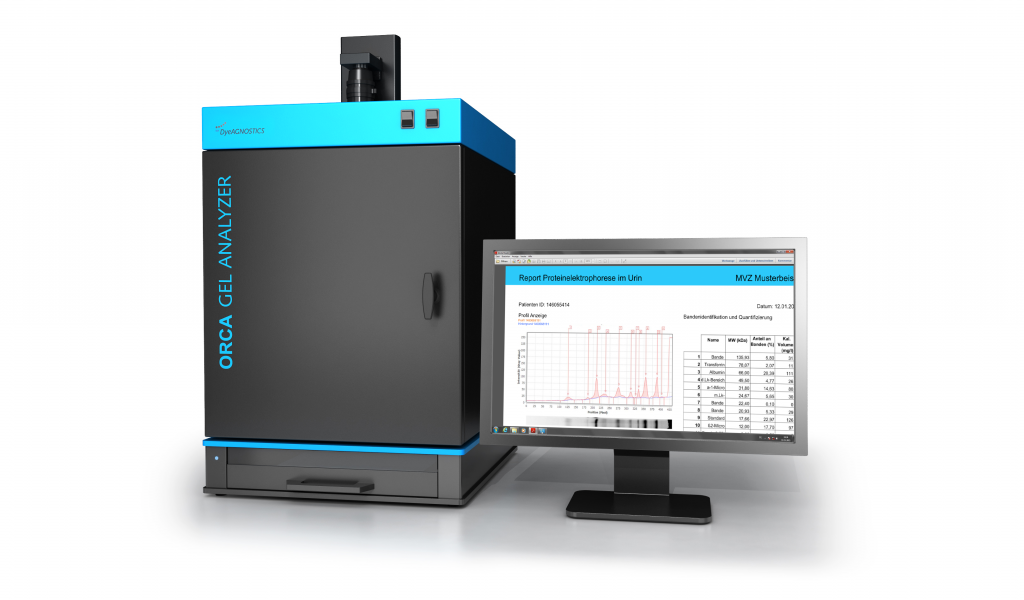
ORCA Gel Analyzer
The ORCA Gel Analyzer - for SDS-PAGE, SAR-PAGE and IEF analysis in daily routine
The ORCA Gel Analyzer provides massive time savings, reproducible results, independency from sample quality (e.g. difference of protein applied to the gel, differences in protein content), digital reporting and independency of the user.
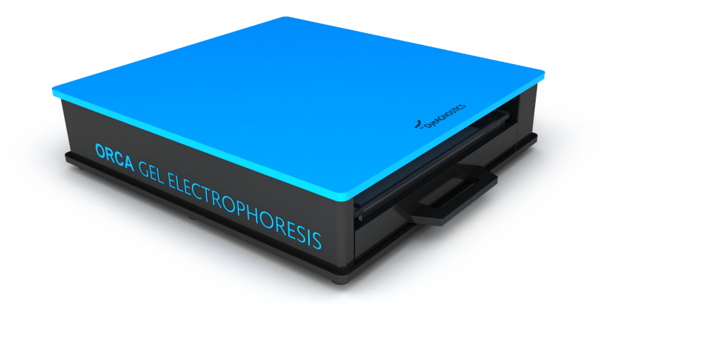

ORCA Gel Electrophoresis Beo Dry blotter
Subcategories
Radioisotope Article Count: 6
Imaging Plates Article Count: 3
Western Blot Article Count: 4